Digital x-rays have become the standard in modern dental practices, yet plenty of patients still wonder whether the technology actually makes a difference or whether it’s marketing dressed up as medicine. The benefits of digital dental x-rays vs traditional film come down to measurable clinical outcomes, not just convenience, and understanding the distinction helps you make a smarter decision about which dental practice deserves your trust and your family’s care.
What You’re Actually Comparing
Traditional film x-rays work the way photography did before digital cameras: a chemical-coated film is exposed to radiation, then processed in a darkroom using developer and fixer solutions. The image appears after several minutes of chemical development. Digital x-rays replace that film with an electronic sensor that captures the same image and transmits it instantly to a computer screen, where it can be viewed, adjusted, and stored in seconds.
The difference isn’t just workflow convenience. The sensor technology and the processing method change the radiation dose required, the quality of the image produced, and the range of things a dentist can actually do with that image once it exists. Every patient benefit that follows from digital x-rays traces back to one of those three changes.
Radiation Exposure
A 2017 systematic review published in the Journal of Evidence-Based Dental Practice examined radiation dose comparisons across x-ray modalities and found that digital sensors require significantly less radiation to produce a diagnostic-quality image than conventional film. A standard bitewing film x-ray delivers approximately 4 to 7 microsieverts of radiation. The digital equivalent delivers roughly 1 to 3 microsieverts, a reduction of 60 to 80 percent depending on the sensor used and the technique.
That reduction sounds abstract until you put it in context. According to the National Council on Radiation Protection, the average American receives about 3,100 microsieverts of background radiation annually from sources like soil, building materials, and cosmic rays at altitude. A single flight from Charlotte to New York exposes you to roughly 6 microsieverts. A digital bitewing falls well below that. A full-mouth film series sits considerably higher. The question to ask at your next appointment is simple: “Do you use digital sensors, and what’s your radiation reduction compared to film?”
How Dose Levels Compare in Practice
A full-mouth series using traditional film delivers approximately 150 microsieverts. The digital equivalent falls closer to 35 to 85 microsieverts depending on the sensor type. That difference may seem small in isolation, but dental x-rays are taken repeatedly over a lifetime. A patient who receives annual bitewings from age 10 through age 70 accumulates a very different lifetime dose depending on whether those 60 years of imaging were captured on film or digitally.
For more detail on how digital sensors reduce cumulative dose, the mechanism comes down to sensor sensitivity: digital receptors respond to lower energy levels, so less radiation is needed to achieve the same image signal.
What This Means for Children and Frequent X-Ray Patients
Children are the patients who benefit most from reduced radiation dose. A 2012 study published in Cancer (Wiley) followed 1,433 patients and found that people who received dental x-rays, particularly bitewings and full-mouth series, at a young age had higher rates of a specific benign brain tumor called meningioma. The study was observational and involved film-era exposures. The clinical takeaway: dose reduction matters most for pediatric patients, and switching to digital x-rays is one of the most direct ways a practice signals that it takes that seriously. Adults requiring monitoring x-rays for periodontal disease, implant follow-up, or orthodontic treatment face similar logic. The dose accumulates, and lower per-visit exposure compounds into a meaningfully safer long-term picture.
Image Quality and Diagnostic Accuracy
A 2020 study published in Dentomaxillofacial Radiology compared diagnostic accuracy between phosphor plate digital receptors and conventional film for detecting proximal caries in extracted teeth. Digital imaging matched or exceeded film accuracy for early-stage decay detection, with the additional advantage that images could be post-processed to improve visibility without re-exposing the patient.
The practical implication: your dentist is more likely to catch a small cavity before it becomes a large one. Early detection consistently means less invasive treatment, lower cost, and less time in the chair.
Enlargement and Enhancement Capabilities
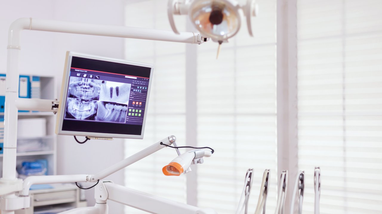
Film produces a static image. Once it’s developed, what you see is what you get. Digital images can be enlarged, adjusted for brightness and contrast, and annotated in real time at the chairside monitor. A shadow near the enamel that’s ambiguous at normal size becomes clearer when enlarged. A subtle change in bone density is easier to track when the previous year’s image loads instantly alongside the current one for direct comparison.
These tools don’t replace clinical judgment, but they extend it. The diagnostic advantage of modern imaging tools lies precisely in giving clinicians more information to act on without requiring additional radiation exposure or additional appointments.
Speed and Chair Time
A 2019 workflow study published in the Journal of Dental Hygiene measured appointment time across film-based and digital x-ray workflows in 22 general practices. Digital practices reduced average x-ray chair time by 11 minutes per appointment when accounting for image development, wet processing, mounting, and darkroom handling that film requires.
For a busy professional or a parent managing a child’s after-school appointment in Charlotte, 11 minutes is meaningful. That’s a difference in whether you make it back for a 2 p.m. meeting or whether your child’s appointment overlaps with a sibling’s pickup. Digital x-rays are available for review within seconds of capture, so the clinical conversation starts sooner and the appointment ends on schedule.
Environmental and Chemical Impact
Traditional film processing requires two primary chemical solutions: developer and fixer. The fixer solution contains silver thiosulfate complexes from the silver halide in the film emulsion. These silver-bearing wastes are classified as hazardous under EPA regulations and require specific disposal protocols. According to the American Dental Association’s environmental guidance, dental practices using film x-rays must use silver recovery units and follow state-specific disposal rules to avoid discharging silver into the water supply.
Digital x-rays eliminate this chemistry entirely. No developer, no fixer, no silver waste, no hazardous disposal contracts. For patients who care about the environmental footprint of their healthcare providers, this is a concrete and verifiable difference, not a marketing claim.
Storage, Accessibility, and Record Portability
Film x-rays are physical objects. They require storage in labeled envelopes, filing systems, and climate-controlled conditions to prevent degradation. Retrieval means locating the physical envelope, pulling it, and mounting it on a lightbox. When a patient transfers to a new practice or needs a specialist referral, the original films either travel with the patient or copies are made, introducing another step and another opportunity for delay or loss.
Digital records are indexed, searchable, and transferable instantly. A general dentist in Charlotte can send a full x-ray series to an oral surgeon or orthodontist the same afternoon, without printing, mailing, or couriering anything.
Sharing Records Across Providers
When treatment involves multiple providers, time matters. An oral surgeon reviewing extraction planning or an orthodontist evaluating jaw development needs current imaging. With film, that process requires physical transport or separate re-exposure. With digital, images move through secure electronic systems the same day a referral is placed. Treatment timelines compress, and patients don’t wait on logistics when they should be waiting on healing.
Patient Communication and Treatment Understanding
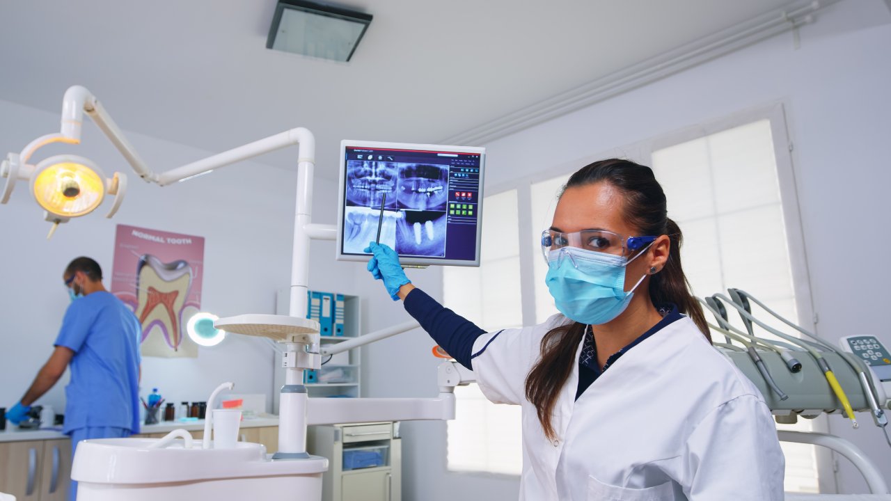
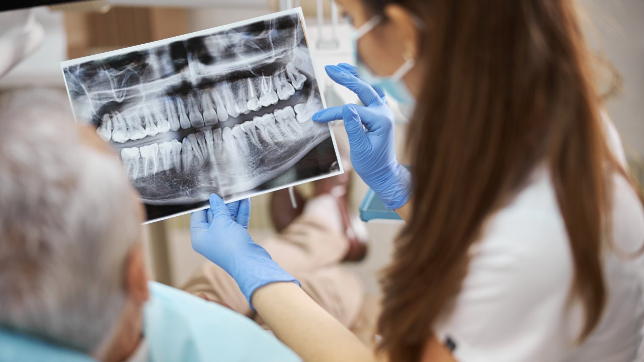
A 2013 study published in the Journal of the American Dental Association found that patients who were shown visual representations of their dental conditions accepted recommended treatment at significantly higher rates than patients who received only verbal explanations. The effect was most pronounced for conditions involving early or moderate decay where no symptoms were present.
When a dentist can display your x-ray on a chairside monitor, point to the exact location of a developing cavity, and show you the size relative to surrounding tooth structure, you stop taking the recommendation on faith and start making an informed decision. That distinction matters. Seeing exactly what your clinical team sees during an examination changes the nature of the conversation from instructions to collaboration. Patients who understand their own conditions make better decisions about treatment timing, and they’re less likely to delay care that shouldn’t be delayed.
Cost Comparison: Digital vs Film
Digital x-ray equipment requires a higher upfront investment than film equipment. A full sensor-based digital x-ray system runs roughly $8,000 to $30,000 per operatory depending on sensor type and software. Film systems are considerably cheaper to purchase initially. That capital cost is absorbed by the practice, and in most cases, it does not translate directly into higher fees for patients. Digital x-ray fees at most general practices are comparable to or lower than film fees, and major dental insurers reimburse digital and film x-rays at the same benefit levels under CDT billing codes.
Long-Term Cost Considerations
Film carries recurring costs that digital doesn’t: film stock, chemical solutions, processor maintenance, darkroom upkeep, and physical storage over decades of patient records. According to practice management research published by the Journal of the American Dental Association, film-based practices spend an estimated $5,000 to $15,000 annually on consumables and processing infrastructure depending on patient volume. Digital practices spend a fraction of that once the initial equipment investment is amortized. Practices that control their overhead more efficiently tend to sustain stable pricing over a long patient relationship, which matters when you’re thinking about a dental home for your family over the next 10 to 20 years.
When Film X-Rays Still Make Sense
Honestly, the honest answer is: rarely, and in narrowing circumstances. Certain specialist applications still use film-based panoramic systems in under-resourced settings where digital equipment hasn’t been acquired. Some forensic dental applications use film because of established evidence protocols. A small number of rural or high-volume low-reimbursement practices have delayed capital investment. None of these scenarios apply to a patient choosing a primary dental home in Charlotte. Film x-rays are not superior to digital in any clinical dimension that affects a patient’s diagnosis, safety, or treatment outcomes.
Which Technology Is Right for Your Dental Care
Families with children benefit most directly from digital x-rays because reduced radiation dose compounds over decades of pediatric and adolescent imaging. Adults managing complex restorative work, periodontal monitoring, or implant follow-up benefit from the comparison and enhancement tools that digital imaging enables. Busy professionals benefit from faster appointments. Seniors requiring regular monitoring benefit from the record portability that makes specialist coordination seamless.
The only patients who aren’t meaningfully affected by the distinction are those who require x-rays rarely, have no complex restorative needs, and never change providers. That describes very few people.
Choosing a Dental Practice in Charlotte Based on X-Ray Technology
When evaluating practices in the Charlotte metro area, ask two specific questions before booking: “Does your practice use digital x-ray sensors?” and “Can I view my x-rays on a screen during the appointment?” Both questions take less than a minute to ask, and the answers tell you a great deal about how a practice thinks about clinical investment and patient communication. A practice that uses digital imaging and displays it chairside has made a specific commitment to transparency. A practice still using film in 2025 has made a different choice, one worth understanding before you make yours.
The Verdict: Digital Wins on Every Clinical Dimension That Matters
The comparison isn’t close. Digital x-rays deliver lower radiation exposure, faster image availability, superior diagnostic resolution, instant record portability, and a chairside communication tool that genuinely changes how patients understand and engage with their own care. Film offers none of those advantages and carries environmental and logistical costs that modern patients shouldn’t have to accept.
The one action to take this week: when you call to book your next dental appointment, ask whether the practice uses digital sensors. If the answer is yes, ask whether they display images at the chair. Those two questions filter for practices that invest in both precision and patient respect, which is the combination that actually produces better outcomes over time.