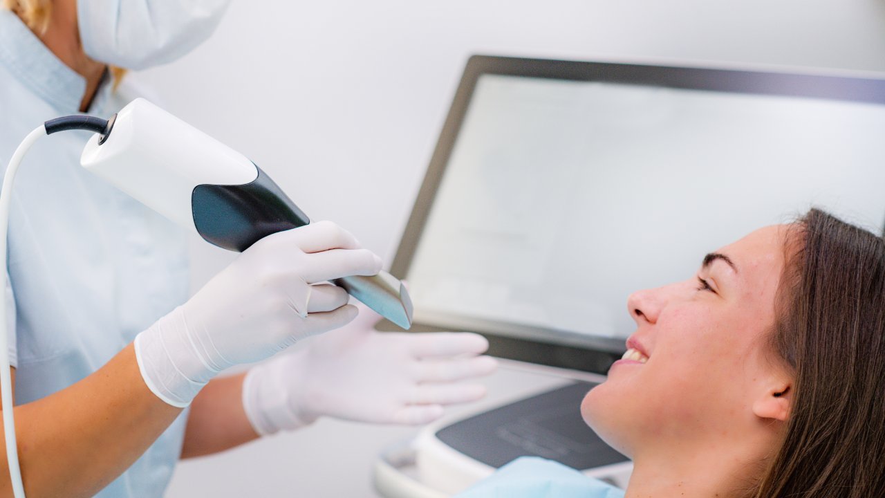
An intraoral camera is a pen-sized wand with a tiny camera tip that captures live, high-resolution images inside your mouth and displays them on a screen you can see from the dental chair. It replaces the old mirror-and-probe guesswork with actual visual evidence. Understanding what an intraoral camera is used for at the dentist changes how you experience every exam and how confidently you can evaluate the dental offices you’re considering.
What Is an Intraoral Camera?
An intraoral camera is roughly the width of a finger and fits comfortably in one hand. The tip rotates to capture angles that no handheld mirror can reach, including the back surfaces of molars, the gumline, and the spaces between older restorations. Images appear on a chairside screen within seconds, in full color, magnified enough to show detail that isn’t visible to the naked eye.
What this replaces matters. The traditional dental exam relies on a small mouth mirror, an explorer probe, and the dentist’s trained interpretation of what those tools feel like against tooth surfaces. That method works, but it leaves a gap between what the dentist perceives and what you, as the patient, can see and understand. The intraoral camera closes that gap completely.
How an Intraoral Camera Works
During the exam, the dentist guides the camera wand around each quadrant of your mouth, pausing at specific teeth to capture still images or short video clips. The tip emits a light source that illuminates the tooth surface and surrounding tissue. There’s no radiation involved, no discomfort from the device itself, and no waiting for film to develop or images to process.
The entire capture process typically adds only a few minutes to a standard exam. Images are saved automatically to your patient file as the dentist works, creating a timestamped visual record without requiring a separate documentation step. The speed is a direct patient benefit: the exam moves efficiently, and the findings are visible before you leave the chair.
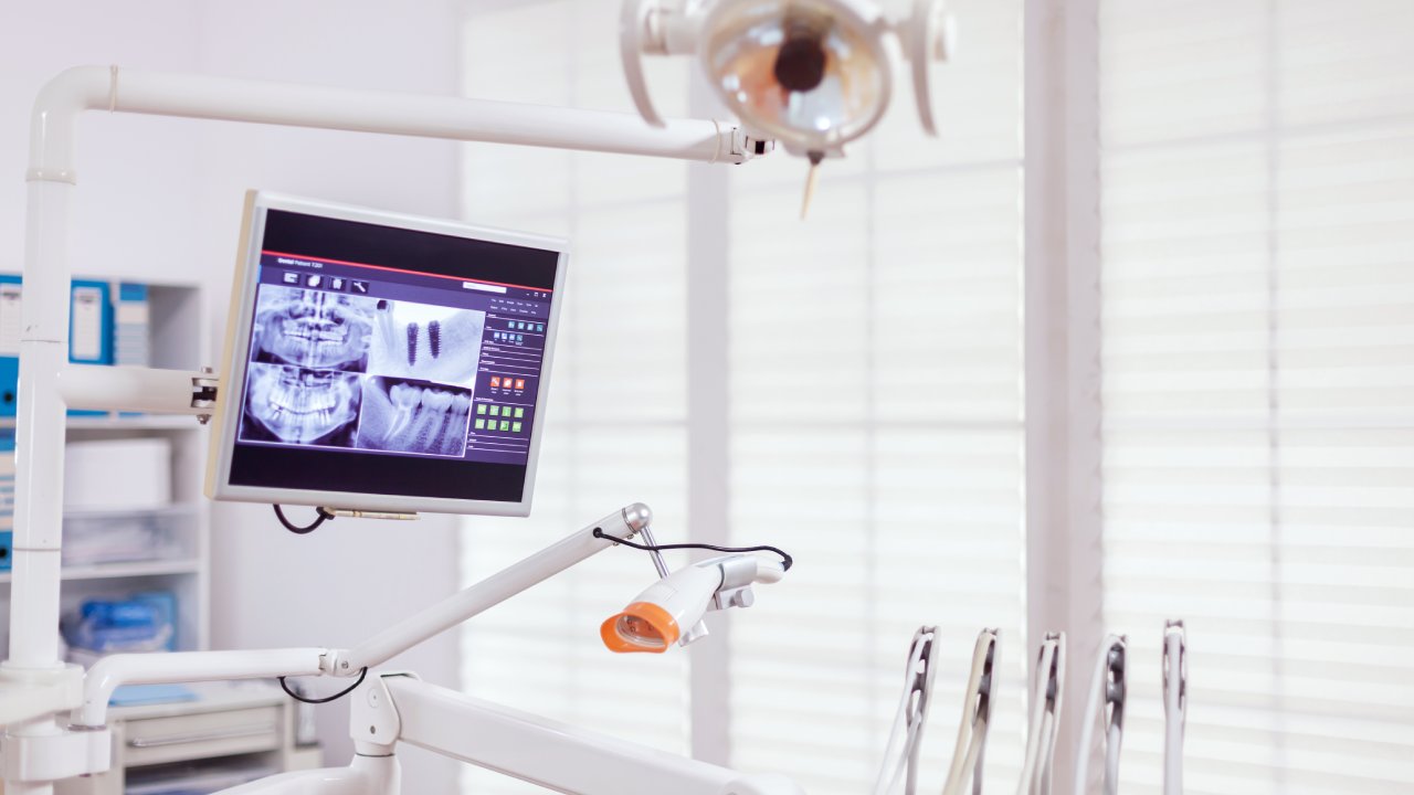
What the Images Actually Show
A 2020 study published in the Journal of Clinical and Diagnostic Research evaluated intraoral camera performance across 320 patients and found that camera-assisted exams detected 34% more early-stage lesions than visual exams alone. The images are that specific.
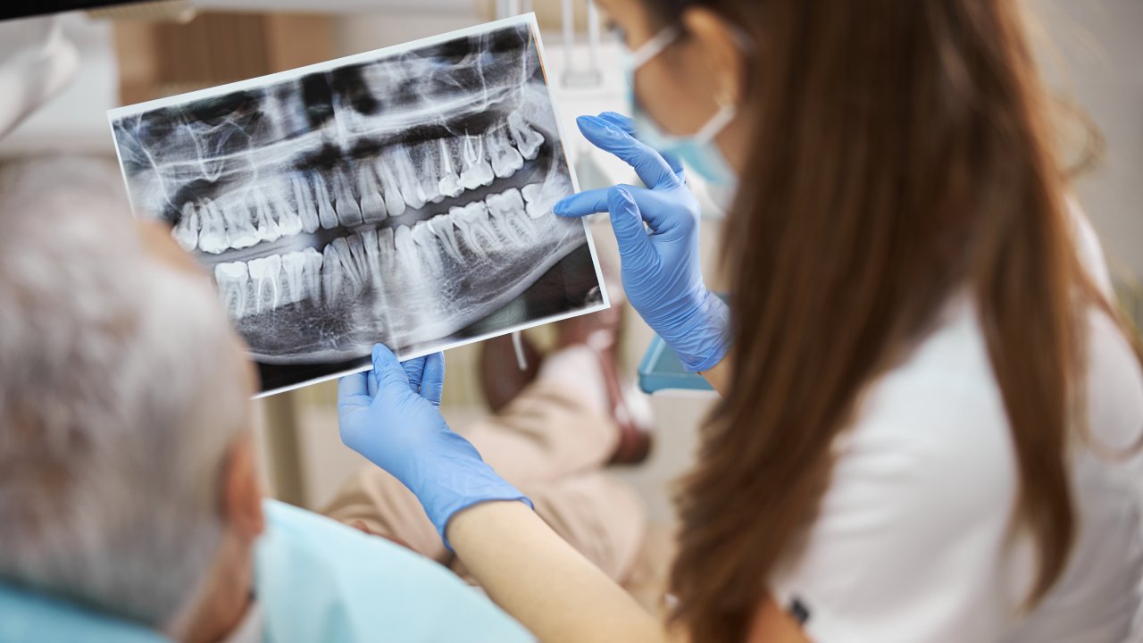
What you see on the screen is the actual surface of your teeth: enamel texture, discoloration patterns, the margins of existing fillings, soft tissue color and contour, and any visible fractures. Compare this to what a standard mirror reveals, which is a small, reversed reflection with limited magnification and no ability to record. The camera doesn’t just help the dentist see better. It lets you see exactly what the clinical team sees, in real time, which is the foundation of any genuinely informed treatment conversation.
What Dentists Diagnose With an Intraoral Camera
The camera’s clinical value shows up most clearly in four areas: cavities and decay, hairline cracks, early gum disease changes, and the condition of existing restorations. Each of these problems shares a common characteristic: they are far easier and less expensive to treat when caught early. The camera’s ability to magnify and illuminate makes earlier detection reliable rather than incidental.
Cavities and Tooth Decay
A 2017 study in the International Journal of Dentistry evaluated 412 patients and found that intraoral camera-assisted diagnosis increased early-stage cavity detection accuracy by 28% compared to visual examination alone. Early-stage decay looks like softened enamel or subtle discoloration, changes that the camera amplifies clearly but that a handheld mirror simply doesn’t show with enough resolution to act on.
What this means in practice: a cavity caught at the enamel softening stage is typically treated with a small filling. The same cavity caught six months later, once it has reached the dentin or pulp, may require a crown or root canal. The camera shifts the clinical window toward the earlier, less invasive, less expensive end of that spectrum.
Cracks and Fractures
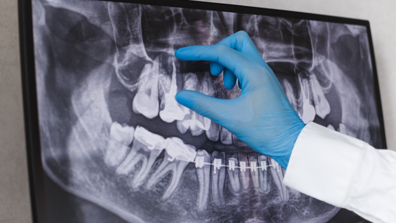
Hairline cracks in teeth are one of the most commonly missed findings in standard dental exams, not because dentists aren’t thorough, but because these fractures are often invisible to the naked eye. A 2019 review in the Journal of Endodontics estimated that cracked tooth syndrome accounts for up to 11% of all tooth loss in adults, with most cases presenting after a crack had already been progressing undetected for months.
Under intraoral camera magnification and the device’s built-in lighting, hairline cracks reflect differently than healthy enamel. They become visible. A crack identified at the hairline stage is typically stabilized with a crown. A crack identified after it has propagated into the root is a candidate for extraction. The camera doesn’t just find what’s already broken. It finds what’s about to break.
Gum Disease and Soft Tissue Changes
According to the CDC’s 2022 periodontal surveillance data, 46% of adults over age 30 in the United States have some form of gum disease. Most of them don’t know it because early periodontal changes, gum recession, tissue inflammation, and early pocketing, are easy to underestimate without documentation.
The intraoral camera creates a photographic baseline. At each visit, the dentist can compare the current image of a gum margin against the image from six months prior. Inflammation that’s stable looks different from inflammation that’s spreading, and a photograph makes that distinction objective rather than interpretive. Understanding how accurate diagnosis shapes treatment outcomes starts here, with documentation that can be measured instead of estimated.
How Intraoral Cameras Support Treatment Planning
A 2021 clinical review published in BMC Oral Health studied 290 patients and found that practices using intraoral cameras to present treatment plans had a 31% higher patient acceptance rate for recommended procedures compared to verbal-only explanations. The mechanism is straightforward: when you can see the problem yourself, the treatment recommendation stops feeling like an opinion and starts feeling like an obvious next step.
Beyond patient acceptance, the camera improves the dentist’s precision in planning. Knowing the exact margins of a crack, the precise extent of decay, or the specific location of gum recession allows the treatment plan to be mapped before any procedure begins. Surprises in the operatory drop significantly when both the dentist and the patient have reviewed the same magnified image beforehand. For anyone evaluating a dental office by its clinical standards, whether a practice uses this kind of shared, image-based planning is a meaningful signal.
The Patient Education Advantage
A 2017 review by Pentapati and Siddiq, published in the National Institutes of Health’s PubMed Central database, examined 14 studies on intraoral camera use and patient compliance. The finding across studies was consistent: patients who were shown intraoral images of their own oral health conditions were significantly more likely to follow through with recommended treatment than patients who received verbal explanations only.
The reason isn’t complicated. Seeing your own molar with a crack visible at the cusp is different from hearing “you have a crack in your molar.” The image makes the problem real and specific. When the dentist shows you the image, ask what the same area looked like at your last visit. That question repositions you from a passive recipient of recommendations to an active participant tracking your own clinical history. It also tells you immediately whether the practice has been documenting your images consistently.
Documentation, Records, and Insurance
Every image captured during an intraoral camera exam is saved to your patient file with a date stamp. Over time, this builds a visual record of your oral health that tracks changes visit to visit and year to year, something no verbal exam note can replicate.
This documentation has practical financial value. A 2020 study in the Journal of the American Dental Association found that insurance claims supported by clinical photographs had a 23% lower dispute rate than claims submitted with written descriptions alone. When your dentist files a claim for a crown or a periodontal procedure, intraoral images showing the condition that necessitated treatment reduce the friction of the approval process.
Ask your dental office to confirm that your images are being saved to your file at every visit, not only when a significant finding appears. A clean baseline image of a healthy tooth is just as valuable as an image of a problem: it establishes what normal looks like for your specific anatomy, which makes future comparisons accurate. Practices that invest in tools that make visits both more precise and more comfortable treat documentation as part of the standard of care, not an optional add-on.
Intraoral Cameras vs. Traditional Dental Exams
The intraoral camera doesn’t replace the dental mirror, the explorer, or X-rays. Each tool has a distinct role. X-rays reveal bone structure, root conditions, and decay that develops between teeth, areas the camera cannot reach. The traditional explorer probe detects texture changes on tooth surfaces through tactile feedback. The intraoral camera adds color, magnification, and real-time imaging of visible surfaces and soft tissue, the layer of clinical information that neither X-rays nor probes can provide.
For a complete diagnostic picture, a well-equipped dental office uses all three. Comparing digital X-rays and traditional film shows a similar pattern: newer formats don’t eliminate older tools, they add a more precise layer to them. Understanding this prevents a common mistake: seeing the camera as a premium feature rather than a standard diagnostic layer. An exam that includes intraoral imaging isn’t a more expensive exam. It’s a more complete one.
What to Try Before Your Next Appointment
When you call to schedule your next cleaning or exam, ask one specific question: does the practice use an intraoral camera, and will the images be reviewed with you at the chairside?
That single question tells you more than most practice websites will. A yes means the office treats diagnosis as a shared process. It also sets the expectation before you arrive, so you enter the appointment ready to engage with what you see rather than waiting passively for a verbal summary. Every visit after that one becomes a data point in a visual record that belongs to you as much as to the practice.