Digital x-rays reduce radiation exposure by a significant, measurable margin compared to traditional film, and understanding how that works helps you make smarter decisions when choosing a dental provider. This isn’t a minor technical upgrade. It’s a shift in how diagnostic imaging operates at a fundamental level, with real consequences for your long-term health.
What Digital X-Rays Are and Why Radiation Dose Matters
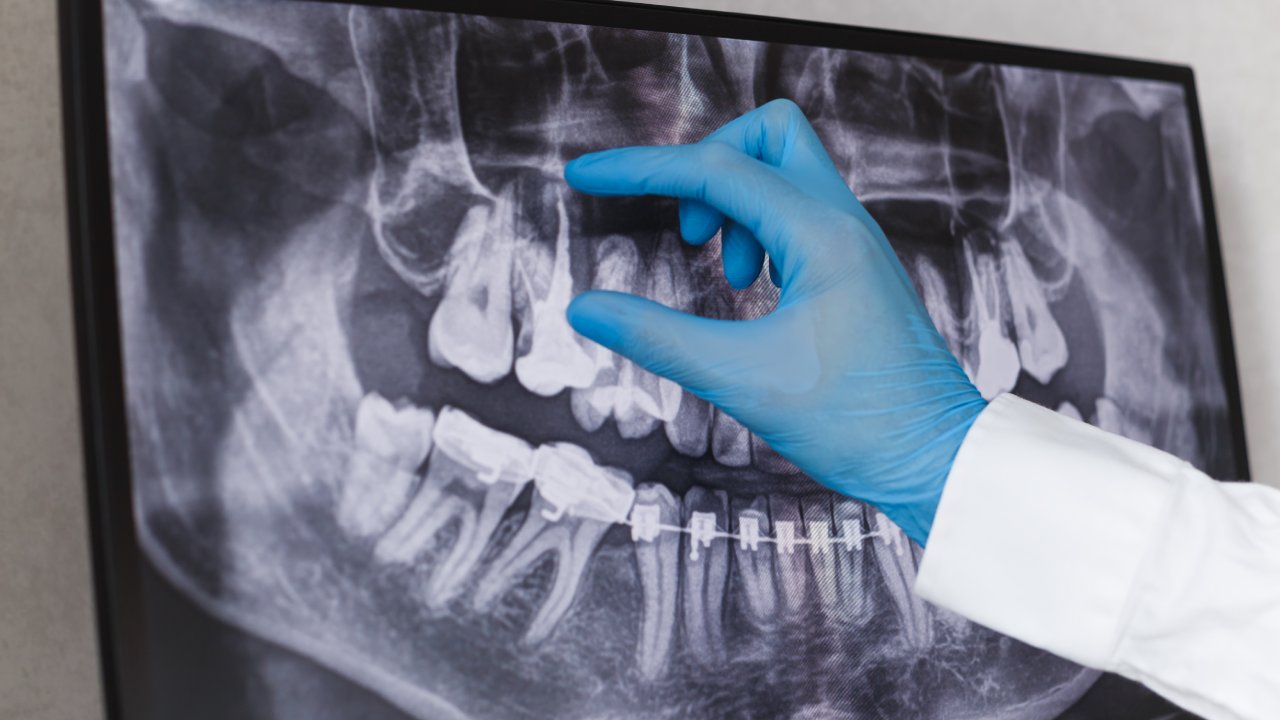
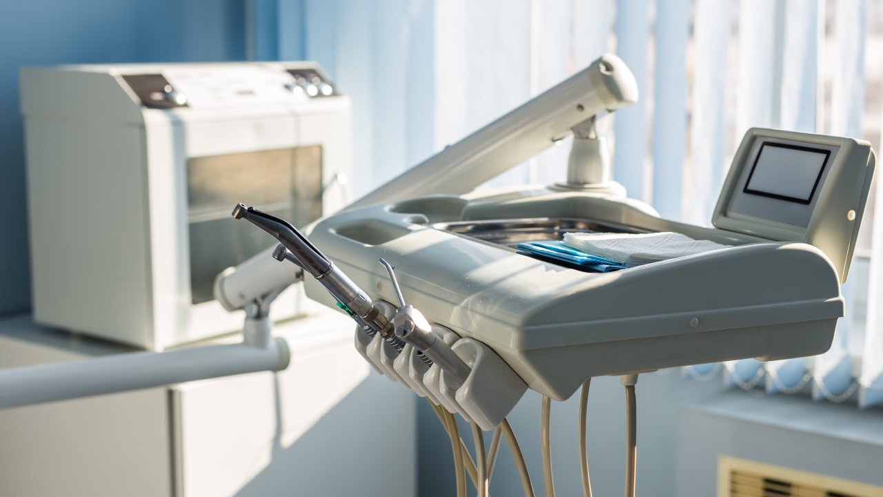
A digital dental x-ray replaces the old plastic film packet in your mouth with an electronic sensor connected to a computer. Instead of waiting for film to be chemically developed in a darkroom, the image appears on a screen within seconds. The diagnostic information is the same. The process is faster, cleaner, and delivers meaningfully less radiation to your jaw, teeth, and surrounding tissue.
Why does radiation dose matter enough to discuss? The FDA has noted that Americans receive more medical and dental radiation exposure than any previous generation, reflecting both longer lifespans and more frequent imaging across all healthcare categories. That cumulative picture is why dose reduction isn’t a minor footnote. It’s a real patient concern, and digital x-ray technology addresses it directly.
How Digital Sensors Capture Images With Less Radiation
The core mechanism is sensitivity. Digital sensors respond to x-ray photons far more efficiently than traditional silver-halide film, which means the beam doesn’t need to stay on as long to produce an image with enough clarity for diagnosis. Less time on means less radiation delivered to your tissues. The relationship is direct.
Research published in the journal Dentomaxillofacial Radiology has confirmed that digital radiography systems require between 50 and 80 percent less radiation than conventional film to produce diagnostically equivalent images. That’s not a marginal gain. For a full-mouth series, the cumulative difference across dozens of exposures is substantial, and it compounds further across a lifetime of routine dental care.
Enhanced Detector Sensitivity
Digital sensors convert x-ray energy into an electronic signal with far greater efficiency than film converts it into a chemical reaction. Film needs a dense exposure to create enough contrast for the dentist to read the image reliably. A digital sensor captures the same information from a fraction of that exposure because the conversion process is simply more efficient at the physics level. The sensor does more work, so the beam does less. That’s the plain-English version of what’s happening.
Reduced Exposure Time in Practice
Exposure times for digital intraoral sensors typically fall between 0.05 and 0.08 seconds per image. Traditional film required exposure times in the range of 0.3 to 0.5 seconds for comparable results, according to figures cited by the American Academy of Oral and Maxillofacial Radiology. At first glance, the difference seems small. But multiply it across a full-mouth series of 18 images and the total dose reduction becomes significant. Across years of dental visits, that difference is one of the clearest arguments for choosing a practice that has invested in current imaging technology when comparing dental offices by technology and quality of care.
How Image Processing Replaces the Need for Repeat Exposures
Traditional film was unforgiving. If the chemical development process produced an image that was too dark, too light, or blurry, the solution was simple and costly: retake the x-ray and double the dose. Digital images appear on screen immediately, and brightness, contrast, and zoom can all be adjusted in software without exposing the patient to a single additional photon.
A 2019 study in the European Journal of Radiology found that digital radiography systems reduced diagnostic retake rates compared to conventional film in dental settings. Fewer retakes means fewer total exposures per appointment. The dose savings from that alone are meaningful, and they accumulate across every visit where something that would have required a retake on film gets corrected with a few clicks instead.
The Role of Collimation and Targeted Beams
Collimation is the process of shaping and narrowing the x-ray beam so it covers only the specific area being imaged, rather than scattering across a wider field of tissue. The smaller the beam footprint, the less surrounding tissue receives any exposure at all.
Digital systems are routinely paired with rectangular collimators, which reduce the exposed field area dramatically compared to the round collimators commonly used with film-based systems. The American Dental Association has identified rectangular collimation as a best-practice dose-reduction tool, and the combination of digital sensors plus rectangular collimation produces the greatest dose reduction of any approach currently in widespread clinical use. If you want to know whether a dental office is serious about limiting your exposure, asking whether they use rectangular collimation alongside digital sensors is a direct, answerable question.
How Digital X-Rays Compare to Everyday Radiation Sources
Context makes the numbers real. A standard digital dental bitewing x-ray delivers approximately 0.005 millisieverts (mSv) of radiation. The National Council on Radiation Protection and Measurements estimates that the average American receives roughly 3.1 mSv per year from natural background radiation alone, just from the environment, soil, and cosmic rays. A cross-country flight delivers about 0.04 mSv. A single digital bitewing is less than what you absorb from a few hours of living on Earth.
The common fear that dental x-rays represent a meaningful radiation risk doesn’t hold up when digital technology is used correctly. The data point in the opposite direction. Understanding why diagnostic imaging matters for clinical accuracy makes it easier to weigh that small exposure against the diagnostic value it delivers.
What ALARA Means and How Your Dental Office Uses It
ALARA stands for As Low As Reasonably Achievable. It’s the governing standard that drives x-ray protocol across dental and medical settings, and it functions as a philosophy as much as a technical rule. Following ALARA means taking x-rays only when they’re clinically justified, using the lowest effective dose settings, and layering dose-reduction tools like digital sensors, rectangular collimation, and protective equipment together.
The FDA and the American Dental Association have published joint guidelines on dental x-ray frequency organized by patient risk category. A low-risk adult with no history of decay and healthy gums doesn’t need a full-mouth series every year. A patient with a history of frequent cavities or active gum disease warrants more frequent imaging. ALARA-compliant practices individualize the schedule based on your actual clinical picture.
If a dental office recommends x-rays at every visit without reference to your history or risk profile, that’s worth a direct question. An office operating under ALARA principles will have a clear answer for why a specific set of x-rays is recommended for you at this appointment.
How Lead Aprons and Thyroid Collars Add a Second Layer of Protection
Lead aprons and thyroid collars block scatter radiation from reaching sensitive tissue during any x-ray exposure, digital or film. The thyroid gland is particularly sensitive to radiation, which is why a thyroid collar that wraps around the neck adds meaningful protection beyond what the primary beam reduction already provides.
The ADA updated its guidelines on thyroid collar use in 2012, and some offices have adjusted their protocols accordingly. The most straightforward step: at your next appointment, confirm that a thyroid collar is offered, especially for children and anyone who is or may be pregnant. A practice that proactively offers both a lead apron and a thyroid collar is demonstrating care at the level of actual clinical detail, not just the headline feature.
Digital X-Rays for Children: Why the Lower Dose Is Especially Significant
Children’s developing tissues are more sensitive to ionizing radiation than adult tissue. The National Academy of Sciences BEIR VII report established this clearly: because children’s cells divide more rapidly during development, they carry a higher sensitivity to the effects of ionizing radiation than adults at comparable exposure levels. That biological reality makes dose reduction in pediatric dentistry particularly important, not just a nice-to-have feature.
Digital x-rays are the standard of care in pediatric dental imaging for exactly this reason. When you’re choosing a dental home for a child, asking whether the practice uses digital radiography is a specific, answerable question that reflects something real about clinical standards. Understanding how current dental technology shapes the patient experience helps you evaluate that answer in context.
The Diagnostic Advantage That Justifies Taking X-Rays at All
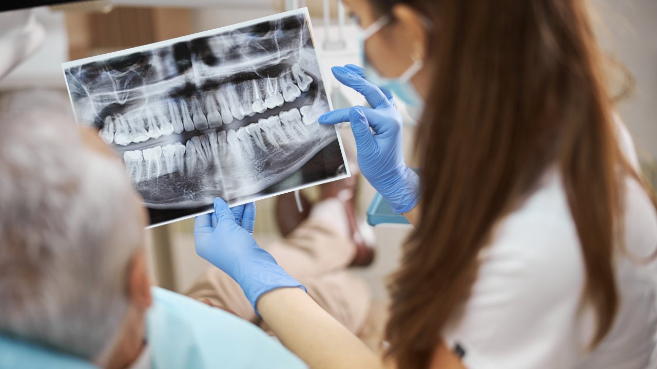
Dose reduction only tells half the story. The reason dental x-rays exist at all is that they catch conditions a clinical exam alone cannot detect: decay developing between teeth, early bone loss, impacted teeth, cysts, and other pathology that stays invisible until it becomes a significantly larger problem.
A study published in the Journal of the American Dental Association found that digital x-rays detect interproximal caries, decay forming between tooth surfaces, at earlier stages than clinical examination alone. Catching decay early means a small filling instead of a root canal, or treatment instead of extraction. The small, measurable dose from a digital x-ray is justified by that diagnostic return, and skipping x-rays out of radiation concern carries its own clinical risk. Missing a cavity for two years because you declined imaging isn’t a conservative choice. It’s a more expensive and more painful outcome.
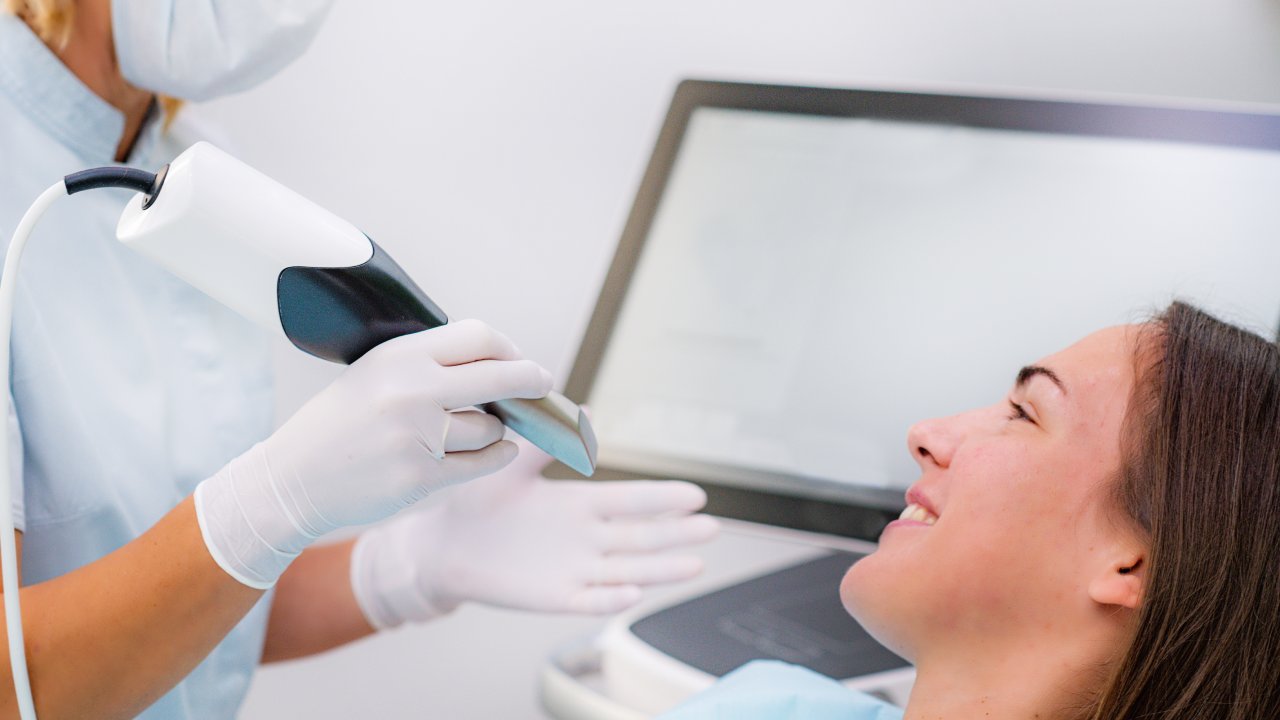
Digital imaging also pairs well with other diagnostic tools like intraoral cameras, which show patients exactly what the clinical team sees in real time, creating a more complete and transparent diagnostic picture overall.
What to Do Before Your Next Dental Appointment
Before you book your next appointment, contact the dental office and ask two questions: do you use digital x-rays, and do you follow ALARA guidelines for x-ray frequency? Both questions take under a minute to answer and immediately tell you whether the practice is operating at the current standard of care. A well-run office will answer both without hesitation and explain how they individualize x-ray schedules based on patient history.
Those two questions work as a practical filter. An office that uses digital radiography and applies ALARA principles has already made the investment in equipment and the commitment to clinical discipline that reduces your exposure while preserving diagnostic accuracy. That combination is the right baseline to expect.