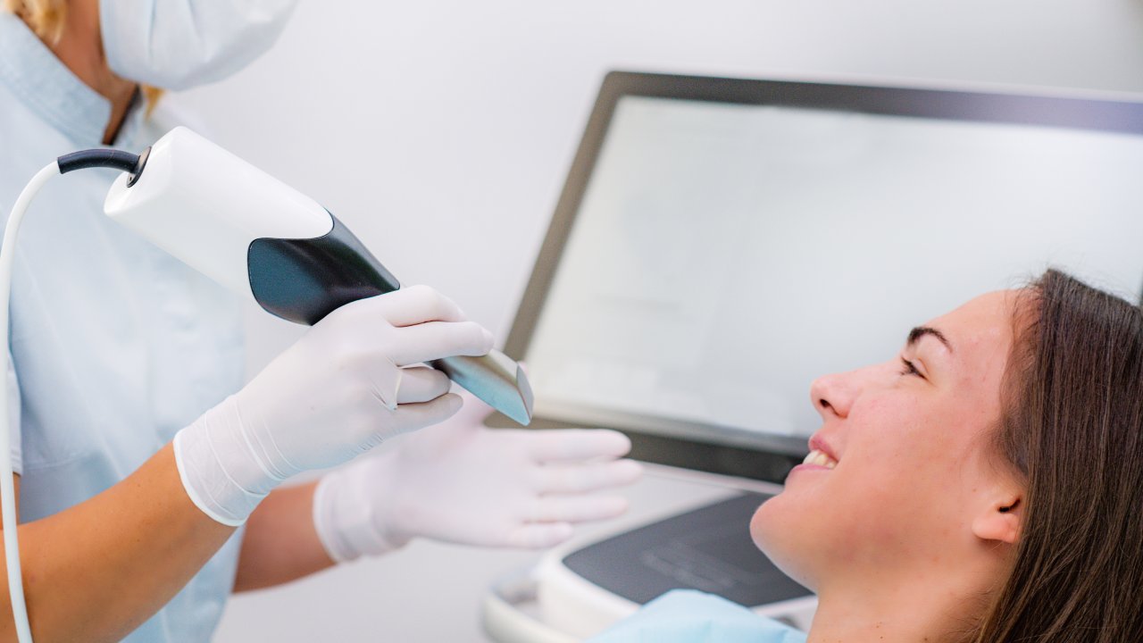
Most patients finish a dental exam knowing something is wrong but not fully understanding what, or why. What an intraoral camera shows patients changes that entirely: it puts a live, magnified view of your own teeth on a screen you can see in real time, turning a professional opinion into something you can evaluate yourself.
What an Intraoral Camera Actually Is
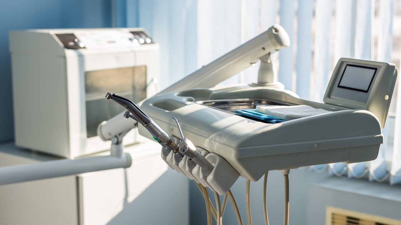
An intraoral camera is a small, pen-shaped device with a lens and LED light at its tip that captures high-resolution video and still images inside your mouth. It is roughly the size of a thick pen, fits comfortably between your teeth and cheeks, and transmits images to a monitor mounted within your line of sight during the exam.
Before this technology existed, the primary visual tool in a dental exam was a small handheld mirror on a metal stick. That mirror showed the dentist a reflected, dimly lit view of one small area at a time. Nothing was recorded, nothing was shareable, and nothing was visible to you. The intraoral camera replaces that limitation with a documented, full-color, magnified record of every surface the dentist examines. That shift from professional-only observation to shared visibility is the reason the technology matters, not the hardware itself.
A Brief History of the Technology
The first commercially available intraoral camera was the ACUCAM system, developed by VideMed and introduced in 1989. Early models were considered a significant clinical advancement, but they came with real practical limitations: bulky equipment carts, cables connecting the wand to a separate processing unit, and image quality that required interpretation even by trained eyes.
Over the following three decades, the technology compressed. Modern intraoral cameras are wireless, produce high-definition images at magnifications that reveal detail invisible to the naked eye, and connect directly to chairside monitors or integrated practice management software. What required a dedicated room and support equipment in 1989 now fits in a drawer and transmits images in under a second. The clinical application, though, has not changed: the camera exists to show both the clinician and the patient what is actually happening inside the mouth, documented in a format that can be stored, compared, and shared.
How an Intraoral Camera Works
The camera emits a focused beam of LED light into the confined, dark spaces of the oral cavity, captures the reflected image through a lens at high magnification, and sends that image to a monitor in real time. The LED lighting is bright enough to illuminate areas that a standard operatory light cannot reach, including the back molars, the interproximal spaces between teeth, and below the gumline margin.
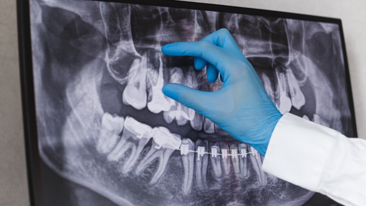
The image the camera produces is a surface-level color photograph. This is worth distinguishing clearly from a dental X-ray, which reveals subsurface structure: bone levels, root anatomy, and decay developing between teeth. A digital X-ray and an intraoral camera answer different diagnostic questions. The X-ray shows you what is happening inside the tooth and beneath the gumline in terms of structure. The camera shows you what is happening on the surfaces you can theoretically see, but with clinical magnification and accurate color reproduction that neither a mirror nor the naked eye provides. For a closer look at how digital imaging compares across modalities, the differences in what each tool captures are meaningful when evaluating any diagnostic finding.
Used together, the two tools give a dentist a complete picture. Neither replaces the other.
What the Camera Actually Shows You
This is where the technology earns its place in a clinical setting. A single exam with an intraoral camera can document conditions across six distinct categories, each of which has direct consequences for treatment decisions.
Early-Stage Tooth Decay
Cavities in their earliest form do not look like holes. They appear as white spots, slight chalky discoloration, or faint shadowing on a tooth surface, changes that are nearly impossible to identify with a handheld mirror under standard lighting. The intraoral camera’s magnification and focused LED illumination make these early lesions visible before they reach the stage that requires drilling.
According to the National Institute of Dental and Craniofacial Research, 92% of adults aged 20 to 64 have had cavities in permanent teeth. Early-stage decay caught on camera can often be remineralized with fluoride or monitored, rather than immediately restored. That is a meaningful clinical and financial difference. Ask the dentist to walk through each image and name what they see: white spot lesion, early demineralization, or established decay. The distinction changes the treatment path entirely.
Cracked or Fractured Teeth
Hairline cracks are among the most common sources of unexplained tooth sensitivity, and they are also among the hardest findings to confirm without magnification. Under the camera’s focused light, a hairline crack shows as a distinct line or shadow across the tooth surface that would be invisible in a standard exam.
The clinical distinction between a craze line, which is a superficial surface fracture that does not threaten the tooth, and a vertical root fracture, which is a serious structural problem, matters enormously for treatment planning. The camera helps differentiate between them based on depth, location, and the light pattern they produce under magnification. If you have sensitivity to cold temperatures or biting pressure, tell the dentist before the exam begins. That information directs where the camera focuses first and speeds up an accurate diagnosis.
Gum Tissue Condition
Healthy gum tissue has a specific appearance on screen: pale pink, firm in contour, with a tight margin at the base of each tooth. Inflamed gum tissue looks different. It is redder, more puffy in contour, and often shows slight bleeding at the margin. Recession, where the gumline has pulled back from the tooth, appears as a visible gap between where the gum should end and where it actually does.
The Centers for Disease Control and Prevention reported that 47.2% of adults over age 30 have some form of periodontal disease. That statistic makes gum assessment one of the most clinically consequential uses of the camera. The redness and puffiness visible on screen correspond directly to bacterial load and inflammatory response at the gumline. Seeing that condition visually, rather than hearing a number called out during a probing exam, makes the finding concrete in a way that changes how seriously patients treat it.
Worn or Damaged Enamel
Enamel erosion from acid exposure, grinding (bruxism), or aggressive brushing technique shows on camera as a flattened occlusal surface, a translucent appearance near the tooth edges, or a worn pattern that reflects light differently than intact enamel. The camera captures this in detail that a standard exam misses.
Enamel does not regenerate. What the camera documents today is a permanent record of a condition that will only progress without intervention. If erosion appears on your images, ask the dentist to pull up images from prior visits and compare. The rate of change, not just the presence of erosion, determines how urgently treatment needs to begin.
Existing Restorations and Their Condition
Fillings, crowns, and veneers do not last forever, and the camera documents exactly where existing restorations stand. Margins where a crown or filling is lifting away from the tooth surface show as a visible gap or dark line. Staining around a restoration margin is an early indicator that the seal has broken and secondary decay is beginning to form underneath.
Secondary decay under a failing restoration is one of the most common reasons patients need more extensive treatment than expected. The camera catches the surface warning signs before the decay spreads beneath the restoration, which means intervention at that stage is still relatively minor. Waiting until there are symptoms typically means the decay has already progressed significantly.
Oral Lesions and Soft Tissue Changes
The camera also documents spots, ulcers, unusual discoloration, or soft tissue changes that warrant monitoring or further evaluation. What distinguishes photographic documentation from a written clinical note is the baseline it creates. An image taken today shows exactly what a lesion looked like at a specific point in time. At your next visit, the dentist compares the new image against that baseline to determine whether the change is stable, resolving, or progressing in a way that requires a biopsy referral.
Written notes like “small aphthous ulcer noted on left buccal mucosa” describe a finding. A photograph of that finding shows it. Those are not equivalent clinical records.
How the Camera Changes the Appointment for You
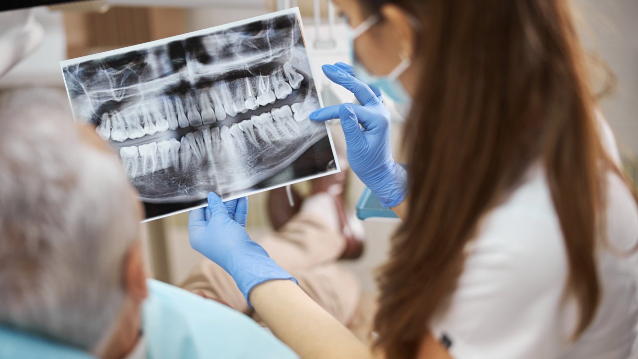
When images appear on a screen you can see in real time, the diagnosis stops being an abstract professional opinion and becomes a visual fact you can evaluate yourself. That shift has measurable consequences for treatment decisions.
A 2017 study published in the Journal of Dental Education found that patients who viewed intraoral photographs accepted treatment recommendations at significantly higher rates than patients who did not, because visual confirmation of a problem removes the hesitation that comes from having to take someone else’s word for it. The plain-language mechanism: seeing it yourself replaces “trust me” with “I can see that.” That is not a small change. It is the difference between a patient who delays necessary treatment because they are not convinced it is urgent and a patient who schedules the follow-up appointment the same day. Understanding why diagnostic transparency matters in clinical settings starts with this kind of direct patient visibility.
The Role of Intraoral Photos in Your Dental Records
Timestamped intraoral images create a longitudinal record of how your teeth change over months and years. That record is clinically valuable in ways that a single visit snapshot is not, because dentistry is largely about monitoring change over time, not just identifying conditions at one point.
Photos document surface and soft tissue changes that X-rays do not capture. If you change dental providers, that photographic history transfers with your records and gives a new dentist immediate visual context rather than a written summary they have to interpret without reference images. Under HIPAA, you have the right to request your records, including photos, and most practices provide them digitally. Request a copy of your intraoral images at each visit and keep them in a personal health file. If a provider cannot or will not provide them, that is worth noting.
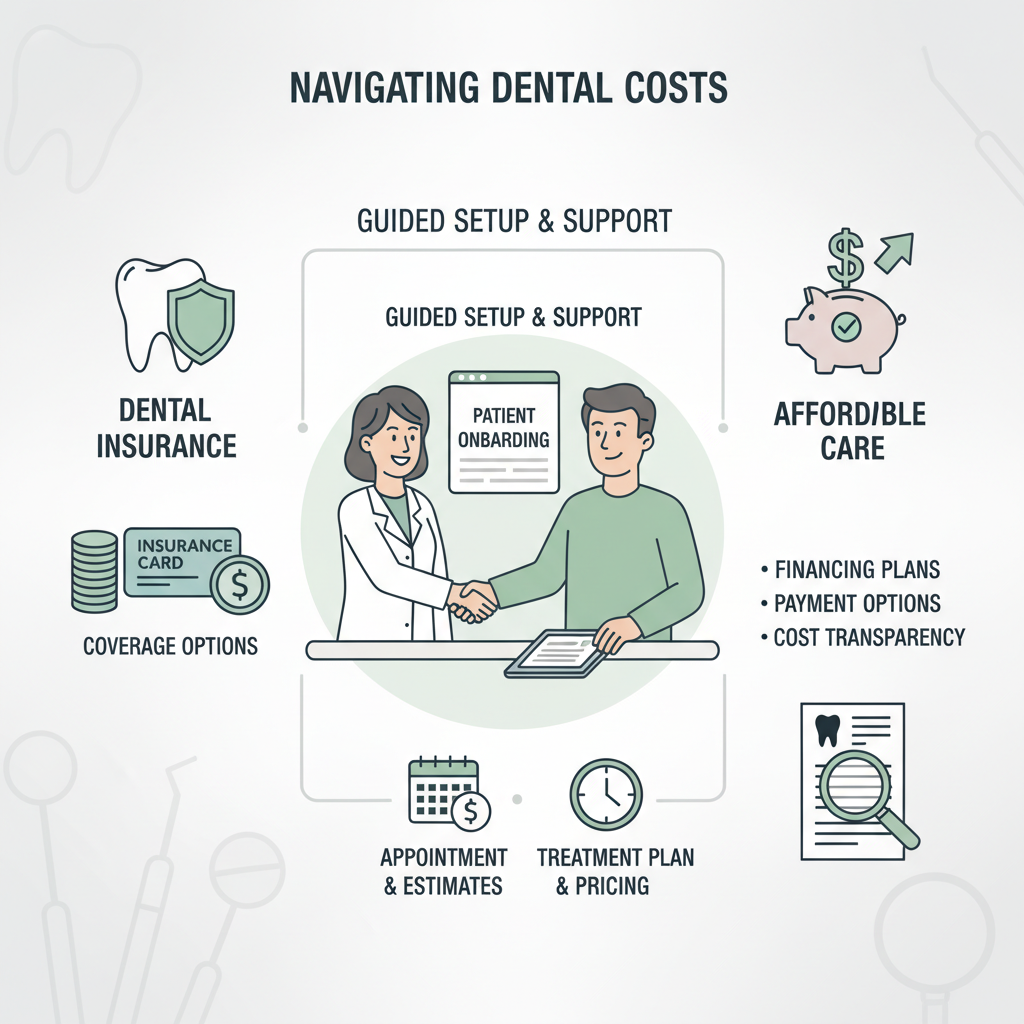
How Intraoral Camera Images Simplify Insurance Claims
Photo documentation provides objective visual evidence that supports pre-authorization for treatment. When an insurer questions why a crown was placed or why a filling was replaced, a dated image showing the crack, the failing margin, or the secondary decay answers that question faster and more definitively than a written clinical narrative alone.
The American Dental Association has noted that claims supported by visual documentation move through the adjudication process more efficiently than those relying solely on dentist notes, because the image removes ambiguity about the clinical finding. Before any major treatment, ask the front desk to note which intraoral images will be submitted with the claim. That one step can reduce back-and-forth with the insurer significantly and accelerates your pre-authorization timeline.
When Dentists Use the Intraoral Camera Most
The camera adds clinical value in several specific contexts, and knowing which appointments involve it helps you know what to expect.
New Patient Exams
A full-mouth series of intraoral photographs at a first visit creates the baseline against which every future exam is measured. A dentist who has no prior images is working without context, evaluating your teeth in a single moment without knowing whether what they see is stable, worsening, or new. That baseline is not a formality. It is the foundation of longitudinal care.
Routine Hygiene Visits
Comparing current images to those from prior visits reveals changes that neither patient nor hygienist would notice visit-to-visit in real time: slow gum recession measured in fractions of a millimeter per year, enamel wear patterns developing on a back molar, early staining at a filling margin that indicates the seal is beginning to fail. This is where the monitoring function of the camera pays off most clearly. When you’re evaluating practices based on how they document and track patient care, the consistency of intraoral photography across routine visits is a meaningful indicator.
Emergency and Diagnostic Appointments
When you arrive with acute pain or a broken tooth, the camera localizes the problem within seconds and allows the dentist to show you the finding before any instrument touches the tooth. That sequencing matters for patient anxiety. Understanding what is happening before treatment begins is consistently associated with lower patient stress during procedures.
What to Expect During the Exam
The wand is moved gently around your mouth while you remain seated. Images appear on the monitor in real time, often positioned where you can see them without turning your head. The dentist or hygienist narrates findings as they capture each image. The exam adds only a few minutes to a standard appointment.
The device is small, the LED light is bright but completely painless, and there is no radiation involved. The camera uses only visible light, and the FDA has cleared intraoral imaging devices for use in clinical dental settings. If you have concerns about the X-ray comparison, there is no comparison to make on safety grounds: the camera produces no ionizing radiation, no heat, and makes no contact with sensitive tissue beyond what a standard instrument would.
The camera does not replace X-rays, and X-rays do not replace the camera. Understanding the specific role of digital X-rays alongside intraoral imaging clarifies why a complete exam uses both, rather than treating them as redundant. X-rays reveal bone levels, root structure, and interproximal decay. The camera reveals surface conditions, soft tissue changes, and restoration integrity. Both are present in a comprehensive exam because neither captures the full diagnostic picture alone.
Every dental office has the option to use this technology, but adoption is not universal. The presence of intraoral cameras in a practice is a reasonable signal that the office invests in diagnostic tools that serve patient understanding, not just clinical efficiency. When you are evaluating a dental home, asking whether intraoral photos are taken at new patient exams and routine hygiene visits is a practical question, not an unreasonable one. Practices that take diagnostic technology seriously as part of patient care treat that question as expected rather than unusual.
What to Ask at Your Next Appointment
At your next dental visit, ask the hygienist or dentist to walk through the intraoral images on screen before discussing any treatment. Name what you see, ask what is being monitored versus what requires action now, and request a digital copy of the images for your records.
That single habit closes the gap between a professional recommendation and an informed decision. A dentist who shows you the crack before recommending a crown is not just being thorough. They are giving you the evidence you need to act with confidence rather than uncertainty. That is the actual value of what an intraoral camera shows patients: not just clinical detail, but the basis for a decision you make with your own eyes.