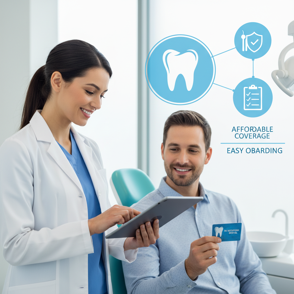
Dental technology isn’t just about having the latest equipment in the room. It’s the single biggest variable between catching a problem at stage one and discovering it at stage four, and that gap has direct consequences for your wallet, your schedule, and how much healthy tooth structure you keep.
The Diagnosis Problem That Older Tools Can’t Solve
A 2019 study published in the Journal of Dental Research found that clinicians using traditional diagnostic methods missed early-stage interproximal caries in roughly 50% of cases on initial examination. That’s not a small margin of error. Decay caught in its earliest stage requires a simple restoration. The same decay caught a year later can mean a root canal, a crown, or an extraction.
The core issue with older diagnostic tools isn’t that dentists using them are careless. It’s that the tools impose a ceiling on what’s detectable. Film X-rays, visual-only exams, and analog impressions all introduce points where precision erodes. What you don’t catch early costs significantly more to treat, in money, chair time, and the loss of tooth structure that can’t be restored once it’s gone. Understanding why dental technology matters for accurate diagnosis starts with understanding those gaps, and how modern tools close them one by one.
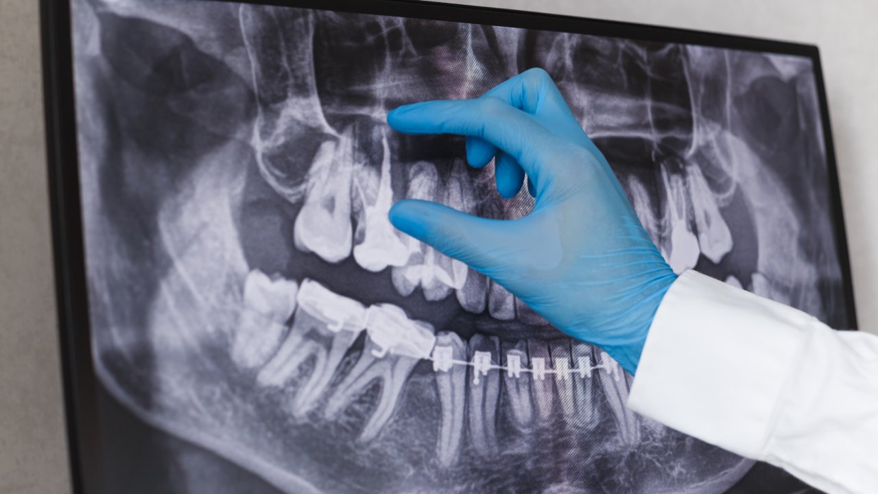
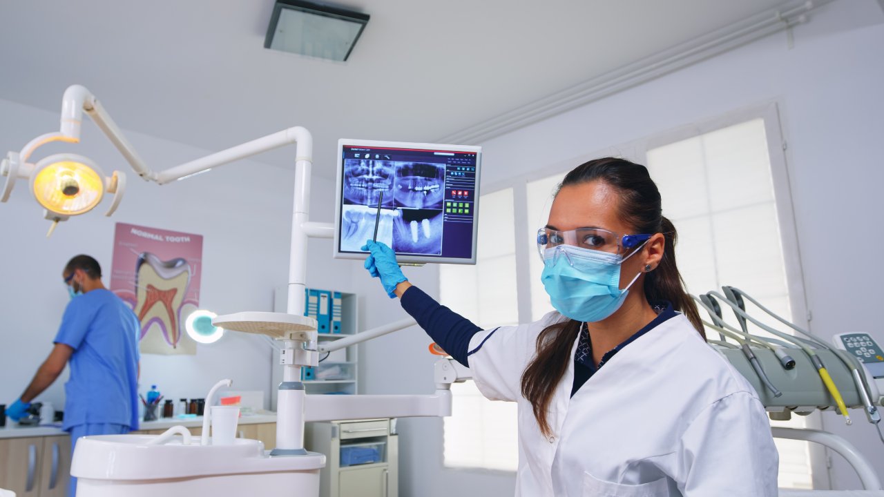
How Digital X-Rays Catch What Film Misses
A 2014 study in Dentomaxillofacial Radiology comparing digital radiography to conventional film found that digital sensors offered superior detection of early proximal caries while delivering approximately 80 to 90 percent less radiation than traditional film techniques. The difference in diagnostic clarity comes from image resolution and the ability to manipulate contrast and brightness after capture, which means a dentist can isolate a shadow on a root surface that would have been invisible or ambiguous on film.
What this means in practice: digital X-rays don’t just produce a safer image, they produce a more useful one. Early bone loss patterns, hairline fractures near the gumline, and decay that hasn’t yet broken through enamel all become readable. If you’re not sure what your dental office uses, that’s a specific question worth asking before your next appointment. The answer tells you something real about the diagnostic depth available to you. For a closer look at how this translates to everyday safety, the breakdown of how radiation compares across imaging types is worth reviewing.
What Lower Radiation Exposure Actually Means for Your Family
For context, a single traditional dental X-ray delivers approximately 0.005 millisieverts of radiation. A digital sensor brings that down to roughly 0.001 millisieverts, which is less than the background radiation you absorb during a single day of normal life. A chest X-ray delivers about 0.1 millisieverts, making a full set of digital dental images roughly equivalent to a few hours of walking around outside.
For parents bringing children in for routine exams, this matters. Children are more sensitive to cumulative radiation exposure than adults, and pediatric dental care often requires more frequent imaging to monitor developing teeth. For seniors managing chronic conditions who see a dentist more often, the same logic applies. With digital equipment in use, the recommended frequency of diagnostic X-rays doesn’t need to be treated as a concern. The radiation reduction isn’t a minor footnote. It’s a structural change in what responsible routine care looks like.
What 3D Cone Beam Imaging Reveals That a Flat Image Can’t
A 2018 systematic review published in Clinical Oral Implants Research evaluated CBCT imaging for implant site assessment and found that 3D imaging altered the treatment plan in 41% of cases where standard 2D radiographs had already been taken. That figure deserves a moment: nearly half the time, the flat image provided an incomplete picture that would have led to a different, and potentially less accurate, clinical decision.
The mechanism is simple. A standard X-ray collapses a three-dimensional structure into two dimensions. Bone density, nerve canal position, sinus floor proximity, and the exact angulation of impacted teeth all require volumetric data to assess accurately. CBCT captures that data in a single rotation, producing a model the dentist can examine from any angle. If you’re being evaluated for an implant, a complex extraction, a TMJ workup, or a root canal on a multi-rooted tooth, 3D imaging isn’t an upgrade. It’s the appropriate standard of care for those cases.
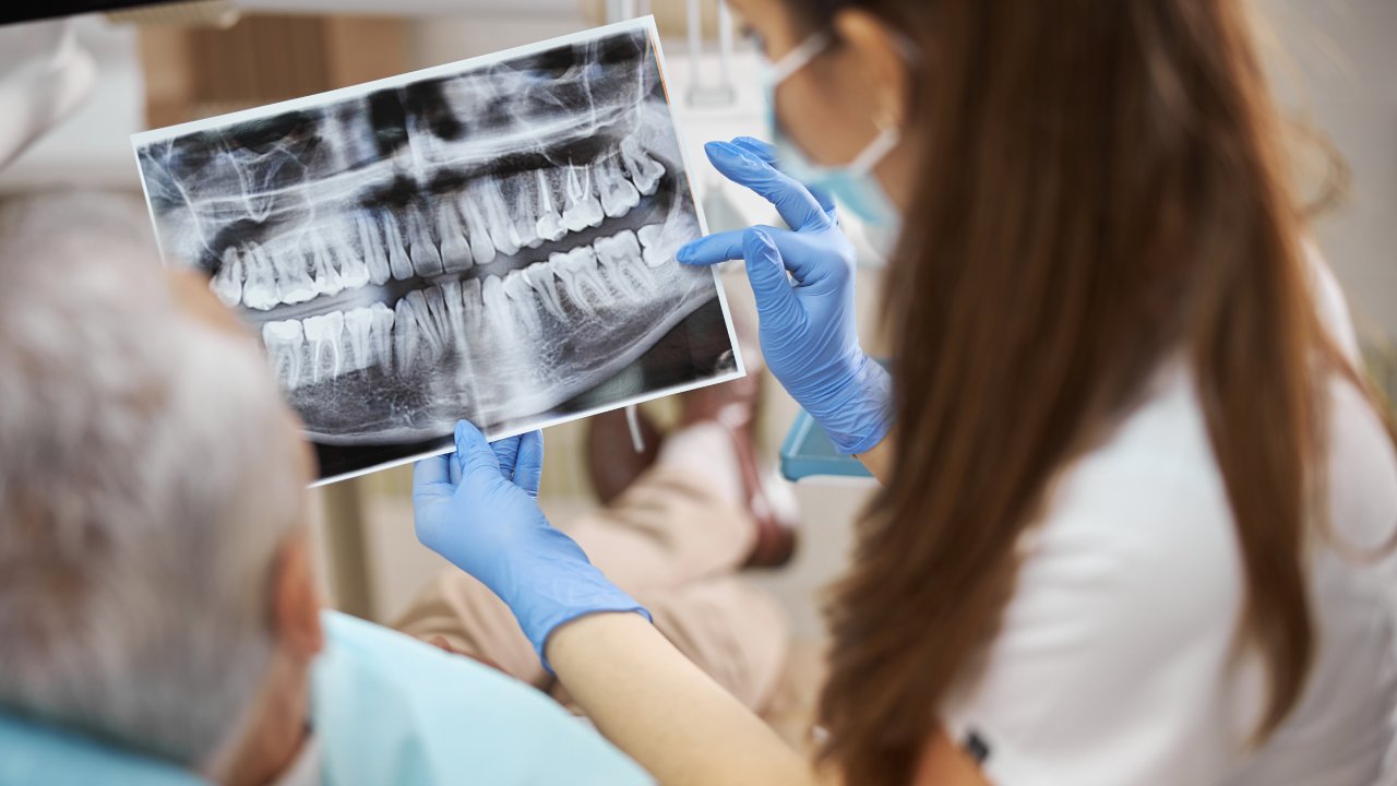
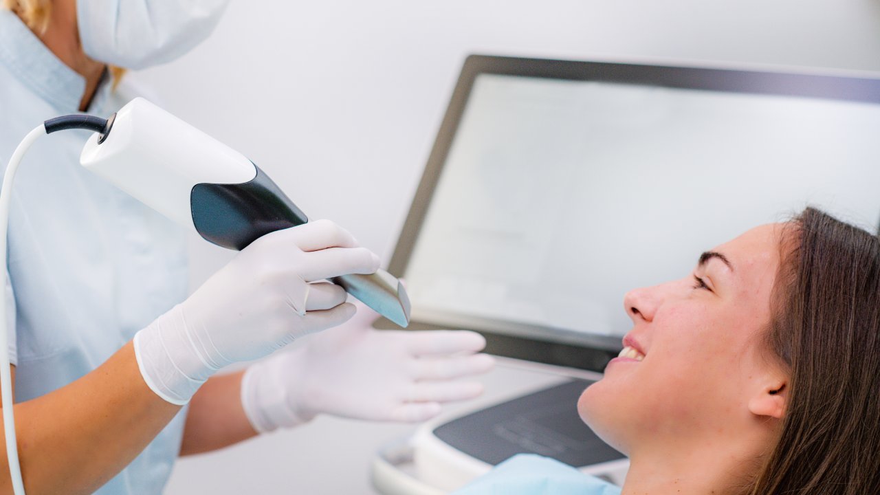
Intraoral Cameras: When You Can See the Problem Yourself
A 2015 study in the Journal of the American Dental Association found that treatment acceptance rates increased significantly when patients could view intraoral camera images of their own teeth compared to verbal-only explanations. The underlying reason isn’t complicated. When a dentist describes a cracked cusp, you’re imagining something. When you see it on a screen, full-color and magnified, the conversation changes.
An intraoral camera is a small wand, roughly the size of a marker, that captures real-time high-definition images of every surface inside your mouth. Cracks that appear only under certain angles of light, early erosion at the gumline, a failing crown margin, a stain that turns out to be decay, all of it becomes visible and documentable. The practical value extends beyond communication. These images create a baseline record of your tooth surfaces over time, which means changes between appointments become measurable rather than impressionistic. During your next exam, ask the dentist to walk you through the images in real time. The ones who use this tool as a communication instrument, not just a documentation tool, will do this readily. For a full picture of what the camera actually shows during an exam, there’s dedicated coverage of how to read those images alongside your provider.
AI-Assisted Detection and What It Adds to a Trained Eye
A 2021 study published in PLOS ONE analyzed AI-assisted radiograph analysis across a dataset of over 3,000 bitewing X-rays and found that AI flagged early-stage interproximal caries with a sensitivity of 87.5%, compared to 75% for unassisted clinician review. That 12-point gap represents real teeth and real treatment decisions.
AI diagnostic tools in dentistry function as a second reviewer. The software analyzes the completed radiograph independently, flagging areas that match learned patterns of decay, bone loss, or lesion formation. It doesn’t replace the dentist’s judgment. It reduces the probability that fatigue, suboptimal film angle, or image noise causes a finding to be missed. The catch is that this tool is only as useful as the practice’s commitment to acting on its output. Ask whether the office uses AI-assisted radiograph analysis. The answer tells you whether the diagnostic workflow includes a systematic check against human error.
How AI Changes the Catch Rate for Early-Stage Decay
The clinical significance of catching decay at stage one versus stage three is a roughly tenfold difference in treatment cost and a complete difference in outcome. Stage one decay, confined to enamel, can sometimes be remineralized without any drilling. Stage three decay, reaching the pulp, requires root canal therapy and a full-coverage restoration. The same lesion. Entirely different paths.
A 2022 review in the Journal of Dentistry found that AI-assisted detection of early caries on bitewing radiographs was 30% more sensitive than standard review alone. What this means in practice: earlier detection leads to less invasive treatment. When you’re comparing dental practices, asking about the diagnostic workflow, specifically whether AI-assisted analysis is integrated, is a direct question about how seriously the practice approaches catching problems before they escalate.
Digital Impressions and the Accuracy Gap in Treatment Planning
A 2017 study in the Journal of Prosthetic Dentistry compared digital intraoral scan accuracy to traditional polyvinyl siloxane impressions and found digital scans produced an average deviation of 49 microns compared to 74 microns for conventional impressions. That difference in precision directly affects how a restoration fits at delivery.
A crown built on an inaccurate impression doesn’t seat correctly. It creates a margin gap where bacteria can infiltrate, changes the bite relationship, and often requires adjustment or remakes. An aligner tray built on a distorted model doesn’t move teeth according to plan. Digital scanning eliminates the dimensional changes that occur when impression material sets, when the model is poured, and when it’s shipped to a laboratory. If you’re scheduled for a crown, implant restoration, or clear aligner treatment, confirming the office uses digital scanning rather than traditional putty molds is a practical pre-appointment question with a direct bearing on what you’ll experience at delivery.
Same-Day Restorations: What CAD/CAM Technology Makes Possible
A 2019 ADA report on chairside CAD/CAM restorations found clinical success rates for same-day ceramic crowns comparable to those produced in a traditional laboratory setting, with patient satisfaction higher primarily due to the elimination of a second appointment and the need for a temporary restoration. The process works as follows: a digital scan feeds directly into milling software, which fabricates a ceramic crown from a pre-shaded block during the same appointment. No temporaries. No second injection of anesthetic. No return visit.
For working professionals in the Charlotte area juggling full calendars, the scheduling difference between one appointment and three is real and quantifiable. One half-day off versus three separate blocks of time, each requiring preparation, transit, and recovery from anesthesia. When a crown is recommended, asking whether same-day CAD/CAM fabrication is available is a straightforward way to determine whether your time and your treatment are both being optimized. To understand the full range of what technology a well-equipped dental practice should have on-site, that resource covers the complete picture.
Laser Dentistry and Precision That Instruments Can’t Match
A 2020 clinical review in Photobiomodulation, Photomedicine, and Laser Surgery found that laser-assisted periodontal treatment produced statistically significant reductions in pocket depth and bacterial load compared to traditional scaling and root planing, with less post-procedural discomfort reported across the study group. The mechanism behind that difference is precision. A laser targets infected tissue with a wavelength specific to its composition, leaving healthy surrounding tissue intact in a way that a blade or scaler cannot replicate.
The diagnostic angle is equally relevant. Laser-assisted soft tissue biopsy produces a cleaner tissue sample with less thermal artifact than conventional excision, which matters when the pathology report needs to be accurate. If gum disease treatment or a suspicious soft tissue finding is on the table, asking whether laser technology is part of the protocol tells you whether you’re getting the most precise available approach to that procedure.
How Technology Compresses the Gap Between Symptom and Solution
A 2021 report from the American Dental Association’s Health Policy Institute found that practices using integrated digital workflows, defined as digital radiography combined with intraoral scanning and AI-assisted analysis, reported 23% fewer unplanned return visits for restoration adjustments compared to practices using conventional methods. Each layer of technology removes a point where error or delay enters the process.
Digital X-rays surface what film misses. CBCT provides the three-dimensional anatomy required for accurate surgical planning. AI analysis provides a systematic review against human fatigue. Intraoral cameras create a shared visual language between patient and provider. Digital impressions eliminate distortion in the model. CAD/CAM fabrication removes the laboratory variable and the second appointment. These aren’t independent features. They’re connected steps in a diagnostic and treatment chain, and the strength of the chain depends on how many links are present. When evaluating dental practices against each other, asking which of these capabilities exist in-office versus requiring an outside referral is the question that separates a practice with genuine diagnostic depth from one relying on a single tool.
What to Do This Week
Call or go online and ask the dental office you’re considering, or currently using, one direct question: which of these capabilities are available in-office? Digital X-rays, intraoral cameras, and 3D imaging represent the diagnostic floor for a practice serious about catching problems early. If three or more of the technologies covered here are absent or referred out, that’s a meaningful signal about the diagnostic depth available to you at that location. Make one call. The information is available and the question is reasonable. That’s the move.