
Benefits of 3D imaging
When you’re planning braces or clear aligners, a high-quality 3d orthodontic imaging service becomes the foundation for precise diagnosis and treatment. These three-dimensional images capture the full complexity of your teeth, jaws, and facial structures. Rather than relying solely on flat X-rays, orthodontists can build a virtual model of your bite, spot hidden issues, and design a treatment plan tailored to your anatomy.
Several key advantages set 3D imaging apart from traditional methods:
- Improved diagnostic accuracy: You and your orthodontist see every root, bone contour, and sinus cavity, reducing guesswork in identifying alignment challenges.
- Personalized treatment planning: Detailed volumetric data let your provider simulate tooth movements, adjust bracket placement for braces, or program clear aligner stages for systems like Invisalign.
- Lower radiation exposure: Modern cone beam computed tomography (CBCT) scanners emit roughly 1.0–3.3 μSv, about 15 times less than a conventional CT scan and comparable to a dozen panoramic radiographs [1].
- Faster turnaround times: Many practices, including Fremont Dental Imaging, offer same-day scan results, helping you start treatment without delay.
By choosing a provider that invests in advanced 3D imaging, you set the stage for a more comfortable experience, shorter treatment duration, and outcomes that align closely with your expectations.
Comparing imaging techniques
Not all orthodontic images are created equal. Understanding the differences helps you ask the right questions and select the best option for your braces or clear aligners.
| Imaging type | Scan time | Radiation dose | Best for |
|---|---|---|---|
| Digital dental X-ray | < 1 second | ~0.005 μSv | Quick checks, cavity detection |
| Panoramic radiograph | 9–12 seconds | ~9–26 μSv | Overall jaw and tooth positioning |
| Cephalometric radiograph | 1–3 seconds | ~5–7 μSv | Side-profile skeletal analysis |
| Cone beam computed tomography (CBCT) | 5–20 seconds | ~1.0–3.3 μSv | 3D jaw, airway, implant and TMJ planning |
Digital dental X-rays
Standard intraoral X-rays produce high-resolution images of a few teeth at a time. They’re indispensable for spotting cavities or checking root health, but they don’t capture broader bone structure or airway dimensions.
Panoramic and cephalometric radiographs
Panoramic images show your full dental arch in a single view, helpful for preliminary alignment checks and wisdom-tooth assessment. Cephalometric radiographs focus on a lateral skull view, allowing your orthodontist to evaluate jaw relationships and growth patterns.
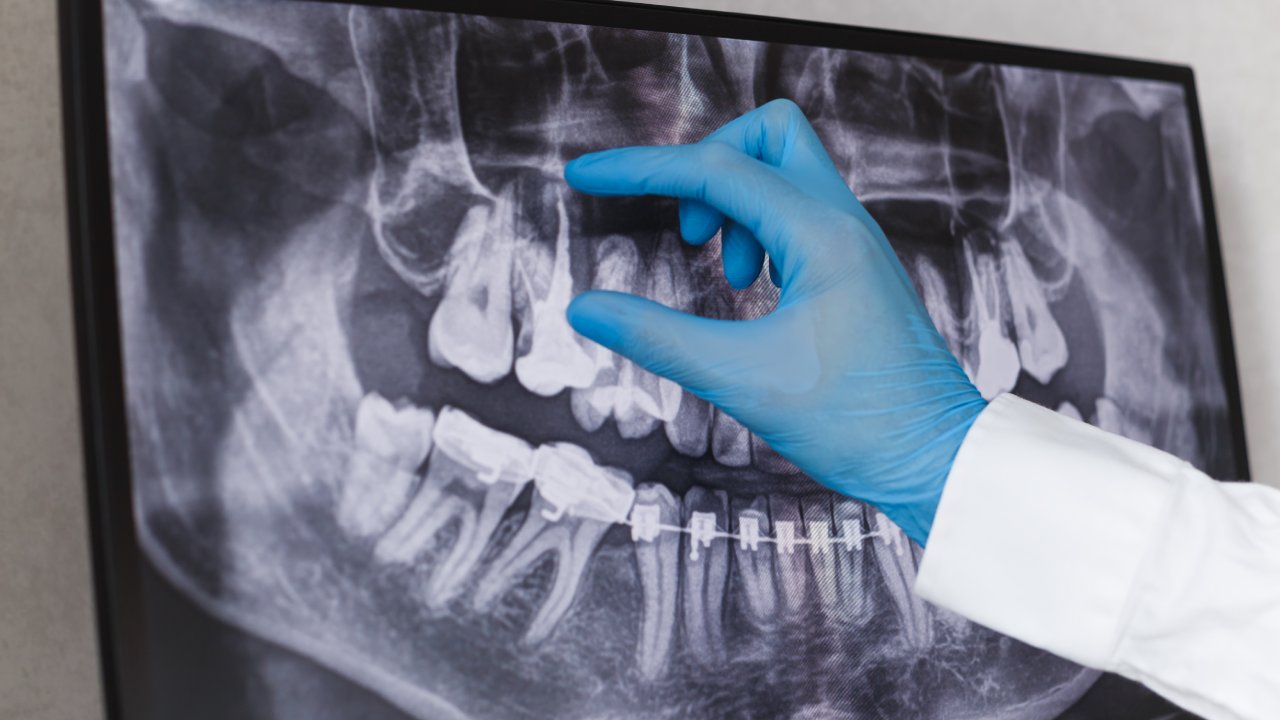
Cone beam computed tomography
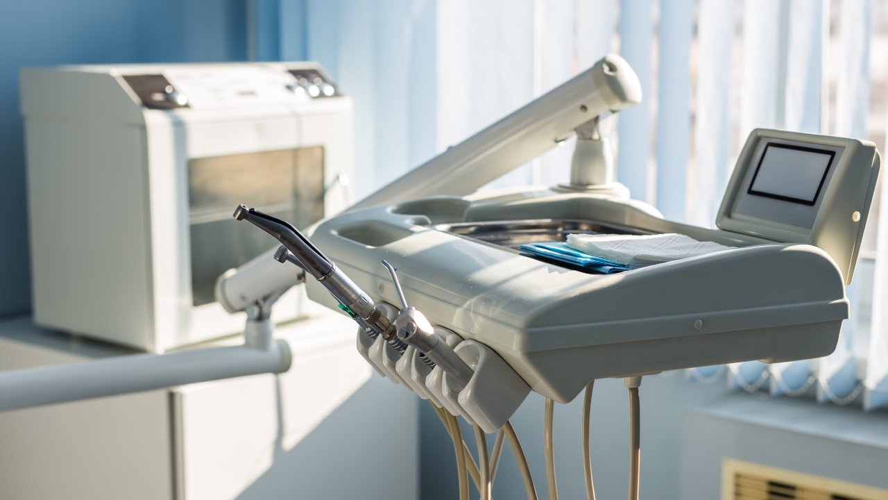
CBCT scanners rotate around your head, capturing hundreds of images in seconds. These slices are reconstructed into a 3D model of hard and soft tissues. Whether you’re considering braces or a clear aligner orthodontic system, CBCT data ensures bracket placement or aligner staging accounts for every nuance of your anatomy. For example, Buffalo’s i-CAT Scan executes a 3D capture in under five seconds with reduced radiation compared to older CT methods [2].
Preparing for your scan
Getting accurate 3D images starts before you ever step into the scanner room. Follow these preparation steps to ensure clear, artifact-free results:
- Schedule an orthodontic consultation. During your orthodontic consultation charlotte, your clinician will determine which imaging study—digital X-ray, panoramic, ceph, or CBCT—best fits your case.
- Remove jewelry and metal objects. Any metal in the head and neck region can cause imaging artifacts that obscure critical details.
- Wear comfortable clothing. Loose collars and hair tied back help you stay still during the scan.
- Arrive on time. Practices like Fremont Dental Imaging confirm pricing and appointment details in advance to keep your visit predictable and efficient [3].
Staying relaxed and motionless for just a few seconds makes a big difference in capturing usable data.
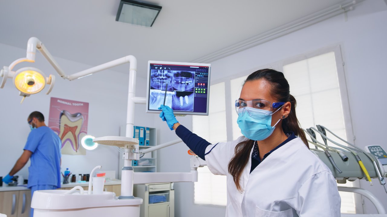
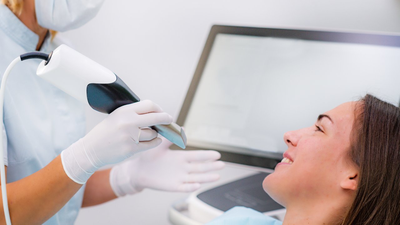
What happens during the scan
When it’s time for your 3D orthodontic imaging service, here’s what you can expect:
You’ll be guided into the scanner and asked to sit or stand in a carefully aligned position. A cone-shaped X-ray beam rotates around your head while the detector moves in unison. Unlike a traditional CT, CBCT scanners capture the entire volume in a single pass, minimizing scan duration.
Keep these pointers in mind:
- The process is painless and non-invasive.
- You may hear a soft whirring noise as the device rotates.
- Total scan time ranges from 5 to 20 seconds, depending on the field of view [4].
- You’ll leave immediately after the scan; there’s no recovery time.
Most providers deliver digital files to your orthodontist within hours or the same day, so your treatment plan can move forward without delay.
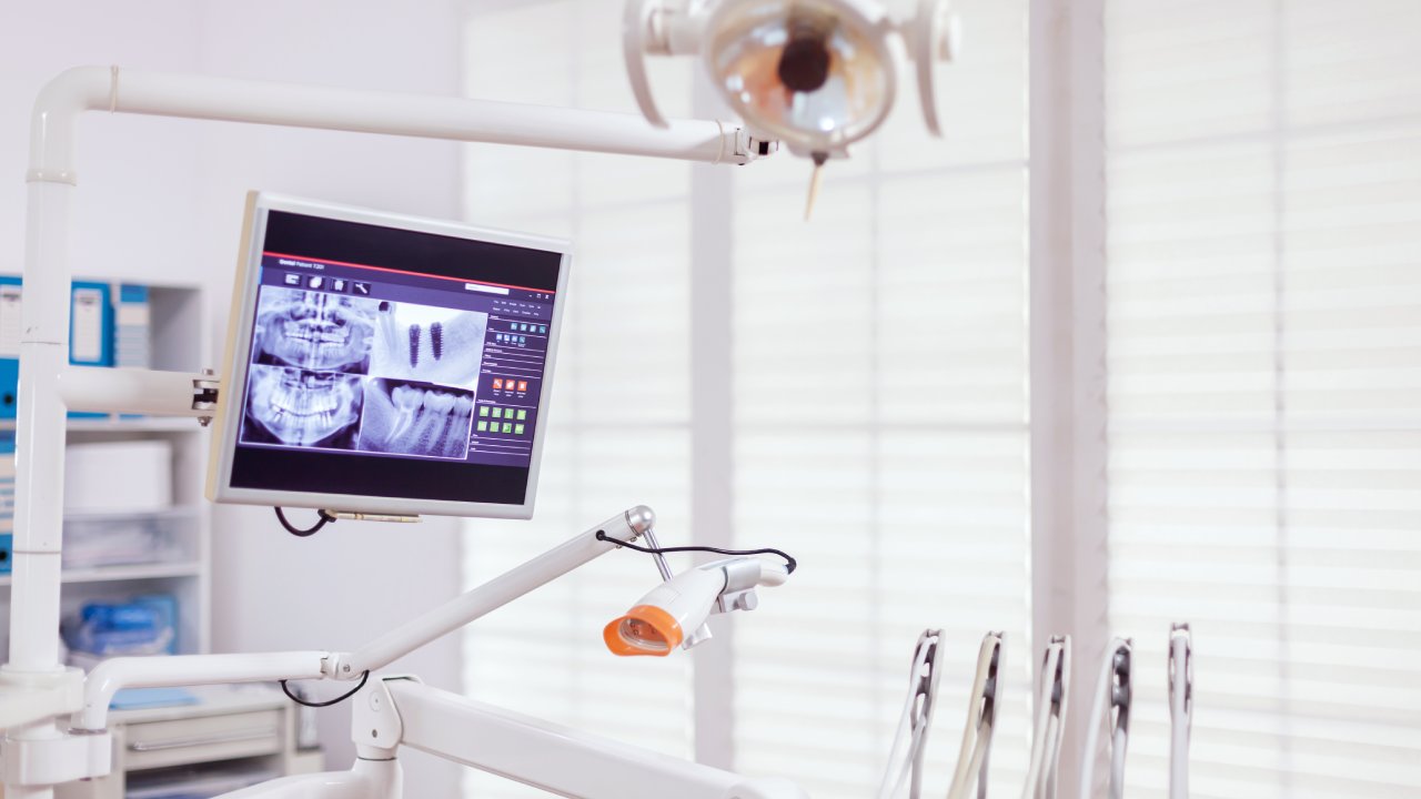
Understanding your results
Once your orthodontist has the 3D dataset, they’ll review cross-sections, volumetric renderings, and measurements to finalize your treatment blueprint. You might see:
- A virtual skull model highlighting crowding, impactions, or asymmetry.
- Precise bone and airway measurements for TMJ or sleep apnea considerations.
- Predicted tooth movements superimposed on your current anatomy.
These insights inform every aspect of your braces or clear aligner plan, from bracket positioning to aligner stage count. By visualizing how teeth will move in 3D space, you gain confidence that the treatment trajectory aligns with your smile goals.
Integrating imaging into braces treatment
The true power of a 3d orthodontic imaging service emerges when those images guide your actual treatment. Whether you choose metal braces, ceramic braces, or an aligner orthodontic system, your orthodontist uses 3D data to optimize outcomes.
Creating a treatment blueprint
Imaging data drives digital treatment planning software. Bracket prescriptions can be customized to your bite forces and root angulations. If you opt for clear aligners—such as Invisalign—you’ll see a simulated sequence of trays that gradually shift teeth into place. Teens can join an Invisalign teen program NC while adults evaluate adult clear aligners treatment.
Tracking progress and adjustments
Follow-up visits rely on periodic scans and photos. Your orthodontist compares new images to the initial 3D model, verifying that tooth movements match the original plan. If a tooth lags behind, digital adjustments can be made to bracket torque or aligner staging. This continuous feedback loop reduces surprises and keeps your treatment on schedule.
Choosing an imaging provider
Selecting the right facility for your 3D orthodontic imaging service ensures accuracy, safety, and value. Focus on two main criteria:
Local options in Charlotte
If you live in Charlotte, look for practices offering dedicated orthodontic imaging. Facilities with experience in dental CBCT understand the nuances of orthodontic cases and collaborate closely with specialists [5]. You want a center that’s comfortable working alongside your orthodontist, whether you’re with a large practice or an independent provider.
Transparent pricing and scheduling
Before booking, confirm fees and policies. Some clinics, like Fremont Dental Imaging, publish USD pricing online and honor those rates unless your clinician changes the requested study [3]. Ask about:
- Cancellation and rescheduling fees
- Insurance billing and out-of-pocket estimates
- Payment options or orthodontic payment plans
A center that clearly explains costs lets you focus on your treatment, not on surprise bills.
After the scan
Your involvement continues once the images are captured. A smooth treatment relies on follow-through.
Follow-up visits and retainer care
After brackets are placed or aligners delivered, you’ll attend regular orthodontic follow up visits. These check-ins allow your provider to review progress, tighten wires, or issue a new set of aligners. At treatment completion, retainer fabrication often uses the final 3D model to guarantee a perfect fit. If your retainer loosens or needs tweaking, a retainer adjustment charlotte visit keeps teeth in alignment.
Monitoring treatment
Occasionally, your orthodontist may request mid-treatment scans to evaluate root parallelism or impacted teeth. Digital imaging also assists in diagnosing and addressing concerns like white-spot lesions or bracket debonding. By leveraging 3D scans throughout your journey, you and your provider maintain a data-driven approach from start to smile finish.
By understanding each stage of the 3D orthodontic imaging service, you’re empowered to make informed decisions—from choosing the right provider to maximizing follow-up care. With advanced imaging guiding every step, your path to a confident, healthy smile becomes clearer and more predictable than ever.